
Continued...
![]()
Continued...
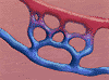 Buds connect with contiguous
buds to form loops. Loops develop a basal membrane from extracellular
matrix components and then develop their own vascular buds. This
process continues until -- Voila! Contact is made with an intact
blood vessel and a capillary loop forms with directed blood
flow. The wound is now adequately re-perfused and this has all been
accomplished within days of the trauma. (Image expandable to 58K
GIF).
Buds connect with contiguous
buds to form loops. Loops develop a basal membrane from extracellular
matrix components and then develop their own vascular buds. This
process continues until -- Voila! Contact is made with an intact
blood vessel and a capillary loop forms with directed blood
flow. The wound is now adequately re-perfused and this has all been
accomplished within days of the trauma. (Image expandable to 58K
GIF).
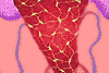
 New highly vascularized
tissue has a granular appearance (which may be why it's called
granulated tissue), and is recognizable by its visible
pinhead-size rounded nodules. Nodules that are dark red in color and
appear moist and shiny indicate good healing. Poor healing is
indicated by a bluish color and a smeary fibrin appearance. (Image
expandable to 36K JPEG).
New highly vascularized
tissue has a granular appearance (which may be why it's called
granulated tissue), and is recognizable by its visible
pinhead-size rounded nodules. Nodules that are dark red in color and
appear moist and shiny indicate good healing. Poor healing is
indicated by a bluish color and a smeary fibrin appearance. (Image
expandable to 36K JPEG).
As the new connective tissue is being created, existing vessels are
recanalized and the old fibrin network is broken down by tissue
plasminogen activator or t-PA. Collagenase, synthesized by
fibroblasts, keeps collagen production in check, and has a critical
function in the process of collagen maturation.
 Part of the process of
collagen maturation includes developing cross-links between
peptide chains or between collagen molecules. It is the cross links
that gives collagen and the scar strength. Also during the maturation
process, collagenase breaks down inappropriately oriented collagen
molecules. The result is that the new collagen, initially laid down
in a chaotic, disorganized way, becomes oriented along the lines of
contour stress in a manner similar to the way nature intended and
nontraumatized collagen appears. (Image expandable to 54K
JPEG).
Part of the process of
collagen maturation includes developing cross-links between
peptide chains or between collagen molecules. It is the cross links
that gives collagen and the scar strength. Also during the maturation
process, collagenase breaks down inappropriately oriented collagen
molecules. The result is that the new collagen, initially laid down
in a chaotic, disorganized way, becomes oriented along the lines of
contour stress in a manner similar to the way nature intended and
nontraumatized collagen appears. (Image expandable to 54K
JPEG).
 Having formed abundant
collagen fibers, the fibroblasts transform either into
fibrocytes or myofibroblasts, the latter has contractile properties.
Consequently the collagen fibers tighten. The end result of the
process? Shrinking scar tissue! (Image expandable to 36K
JPEG)
Having formed abundant
collagen fibers, the fibroblasts transform either into
fibrocytes or myofibroblasts, the latter has contractile properties.
Consequently the collagen fibers tighten. The end result of the
process? Shrinking scar tissue! (Image expandable to 36K
JPEG)
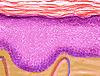 Ah . . .but the wound
surface, you wonder. Repair of the epithelium is needed to
keep the undesirables out and the vital essentials -- body fluids and
electrolytes, for example -- from escaping. Reconstruction of the
epithelium of the skin is a Herculean task as the epidermis is
composed of four layers: the basal layer, which lies just above the
dermis, the prickle cell layer, the granular layer, and finally the
stratum corneum, which is composed of dead cells and keratin.
(Image expandable to 46K JPEG).
Ah . . .but the wound
surface, you wonder. Repair of the epithelium is needed to
keep the undesirables out and the vital essentials -- body fluids and
electrolytes, for example -- from escaping. Reconstruction of the
epithelium of the skin is a Herculean task as the epidermis is
composed of four layers: the basal layer, which lies just above the
dermis, the prickle cell layer, the granular layer, and finally the
stratum corneum, which is composed of dead cells and keratin.
(Image expandable to 46K JPEG).
 Reconstruction takes place in
four stages: Detachment, migration, proliferation, and
differentiation. Within 24 hours of wounding, the basal cell layer of
the epidermis adjacent to the wound thickens and marginal cells
elongate and migrate over the wound. The cells at the advancing
epithelial edge are followed by a monolayer of new epithelial cells.
The new epithelial cells advance across the wound until they
eventually meet epithelial cells moving in from the opposite
direction. (Think of it as being like the meeting of the
transcontinental railroad.) (Click on image to download Quicktime
movie)
Reconstruction takes place in
four stages: Detachment, migration, proliferation, and
differentiation. Within 24 hours of wounding, the basal cell layer of
the epidermis adjacent to the wound thickens and marginal cells
elongate and migrate over the wound. The cells at the advancing
epithelial edge are followed by a monolayer of new epithelial cells.
The new epithelial cells advance across the wound until they
eventually meet epithelial cells moving in from the opposite
direction. (Think of it as being like the meeting of the
transcontinental railroad.) (Click on image to download Quicktime
movie)
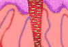
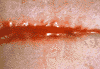 The
price of wound repair is a scar, which appears reddish at
first but as the connective tissue grows tauter and vascularization
slows, it gradually looses color. Hair, sebaceous and sweat glands
are also absent, as is the ridged pattern of the epidermis. Thus the
new skin's appearance is unusually smooth. (Image expandable to
36K JPEG)
The
price of wound repair is a scar, which appears reddish at
first but as the connective tissue grows tauter and vascularization
slows, it gradually looses color. Hair, sebaceous and sweat glands
are also absent, as is the ridged pattern of the epidermis. Thus the
new skin's appearance is unusually smooth. (Image expandable to
36K JPEG)
 Go back to first page of New Tissue Formation Go back to first page of New Tissue Formation |
Please see Wound Healing in Specific Tissues |
Home | Welcome | Clinical Update | Managing Your Residency | Board Review | Opportunities | Links | Contributors | Contact Us